Understanding this service
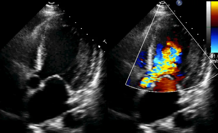
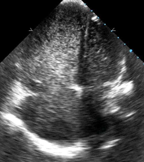
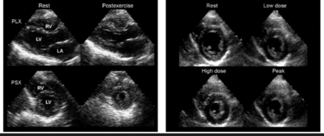
Echocardiography uses high-frequency sound waves to create real-time images of the heart. A standard transthoracic echocardiogram is performed from outside the chest and shows the size and movement of the chambers, the heart muscle, valves and patterns of blood flow.
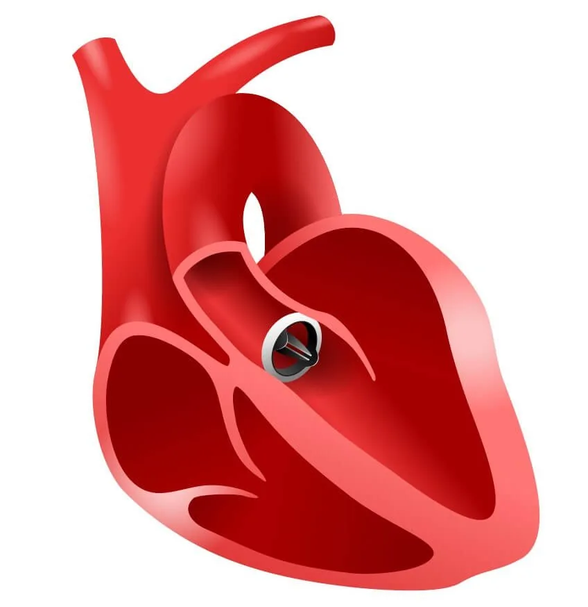
The source heart-valve page identifies echocardiography as the main test for finding the cause of valve disease, measuring severity and choosing treatment. It is also widely used to assess heart function, heart failure, murmurs and structural conditions.
When is it recommended?
- Breathlessness, swelling, fatigue or symptoms that may reflect reduced heart function
- A heart murmur or suspected valve narrowing or leakage
- Assessment after a heart attack or in known coronary heart disease
- Suspected cardiomyopathy, congenital heart disease or fluid around the heart
- Follow-up of known heart-muscle, valve or structural disease
Purpose and potential benefits
- Shows heart chambers, muscle movement and overall pumping function
- Assesses valve structure, narrowing, leakage and blood-flow patterns
- Uses ultrasound rather than ionising radiation
- Provides measurements that support diagnosis, treatment planning and follow-up