Understanding this service
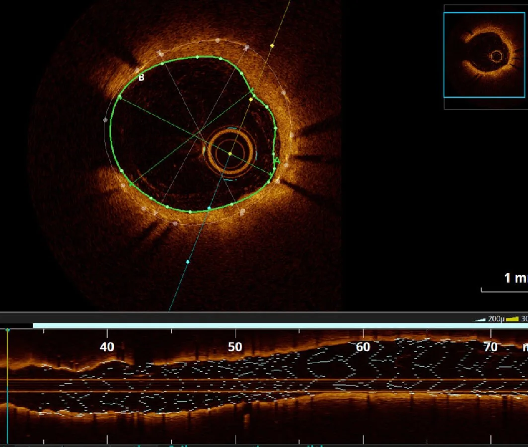
Intravascular imaging uses a very small imaging catheter inside a coronary artery to show the vessel wall and lumen in greater detail than angiography alone. Common techniques include intravascular ultrasound (IVUS) and optical coherence tomography (OCT).
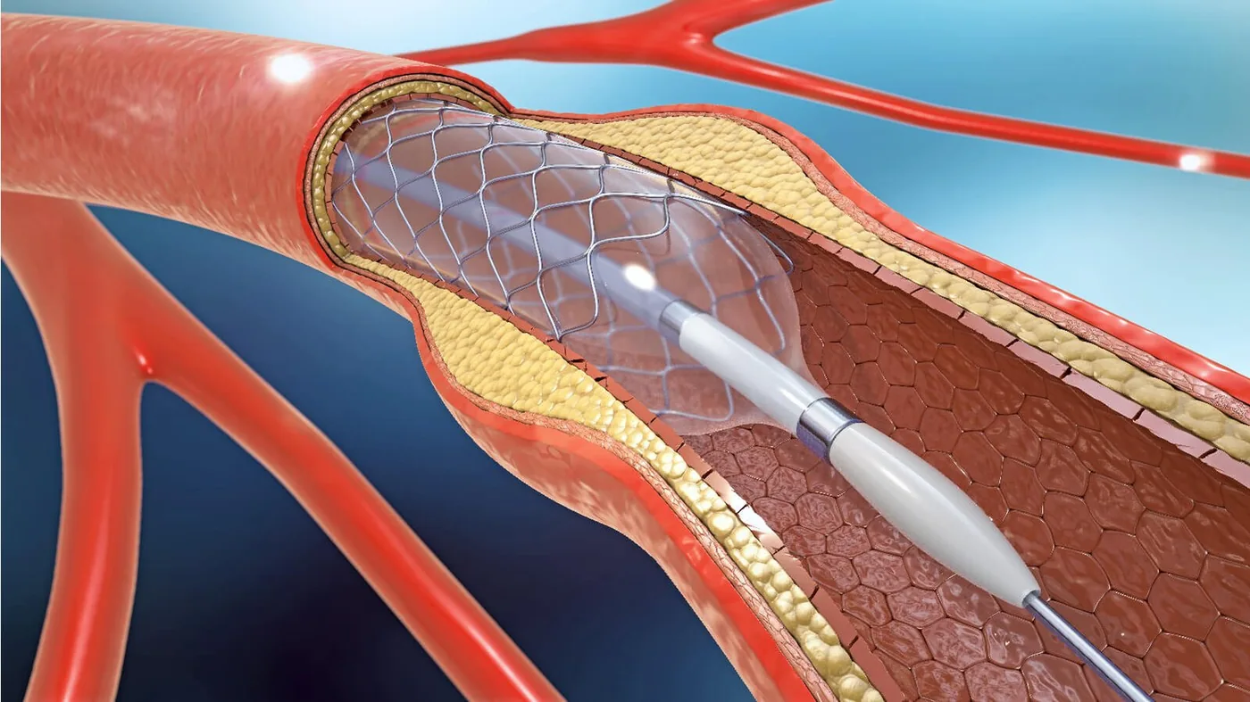
It is usually performed during coronary angiography or angioplasty rather than as a separate operation. The images can help the cardiologist understand a lesion, measure the artery and assess how well a stent has expanded and contacted the vessel wall.
When is it recommended?
- A coronary narrowing that is difficult to assess fully on angiography alone
- Planning selected complex angioplasty procedures
- Choosing an appropriate stent size and landing zone
- Checking stent expansion, position and contact with the artery wall
- Investigating selected cases of recurrent narrowing or a problem within a stent
Purpose and potential benefits
- Provides a cross-sectional view from inside the coronary artery
- Adds vessel measurements that can support treatment planning
- Helps identify features that may not be clear on a two-dimensional angiogram
- Allows the cardiologist to assess and optimise a newly placed stent