Understanding this service
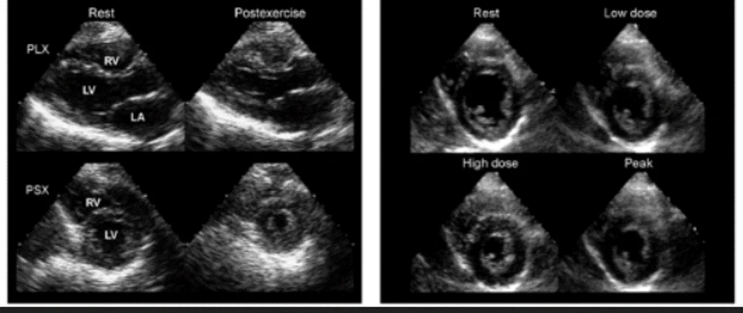
Stress echocardiography combines ultrasound imaging with controlled physical or medication-induced stress. Images taken at rest are compared with images obtained when the heart is working harder.
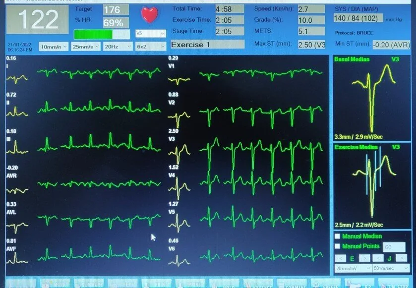
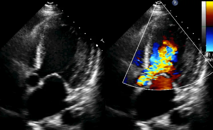
The test can assess heart-muscle movement, pumping response and selected valve conditions under stress. Exercise is commonly used when possible; medication may be used when a patient cannot exercise adequately.
When is it recommended?
- Symptoms that occur with exertion and require imaging beyond an exercise ECG
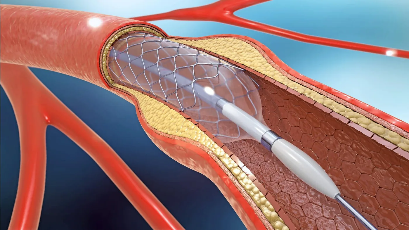
- Assessment for reduced blood flow to part of the heart muscle
- A resting ECG that makes a standard treadmill test harder to interpret
- Evaluation of exercise-related changes in selected valve disease
- Follow-up or risk assessment in selected people with known coronary disease
Purpose and potential benefits
- Compares heart-wall movement and pumping function at rest and under stress
- Adds ultrasound images to heart-rate, blood-pressure and symptom monitoring
- Can evaluate functional effects that may not appear at rest
- Does not use ionising radiation